
CTA vs MRA: Which One Should You Choose?
Kate Williamson, Editorial Team, European Hospital & Healthcare Management
CTA and MRA are both non-invasive imaging tests for visualizing blood vessels, but they differ significantly. CTA uses X-rays and iodine contrast, offering speed and sharp detail, ideal for emergencies. MRA uses magnetic fields, avoiding radiation, better for repeated monitoring. Choice depends on urgency, implants, kidney function, and specific vascular concerns.
Introduction:
If your doctor has just told you that you need an "angiography," there's a good chance you've already run into a wall of confusing acronyms. CTA. MRA. MRI angiography. CT angiography. They all sound similar, they all look at your blood vessels, and yet they are not the same test at all.
This guide breaks down CTA vs MRA in plain language — what each scan actually does, how they differ, and how doctors decide which one fits your situation. Whether you're prepping for an appointment or just trying to understand a report you were handed, you'll walk away knowing exactly what separates these two tests.
What Is CTA (CT Angiography)?
CT angiography is a scan that combines a standard CT (computed tomography) machine with an injected contrast dye to create detailed, 3D images of your blood vessels. The machine uses X-rays, captured from multiple angles, then a computer stitches those images together into cross-sectional "slices" — and eventually a full 3D map of your arteries or veins.
How it works:
- A contrast dye (usually iodine-based) is injected into a vein
- The CT scanner takes rapid X-ray images as the dye moves through your blood vessels
- Software reconstructs the images into detailed 2D and 3D views
- What it's good at:
- Speed — a CTA scan often takes just a few minutes
- Sharp detail on calcium deposits and plaque buildup
- Excellent for emergency situations (like suspected pulmonary embolism or aortic dissection) because it's fast and widely available
- Strong performance in patients with implanted metal devices like pacemakers
- What Is MRA (MR Angiography)?
MR angiography uses magnetic fields and radio waves — not X-rays — to produce images of your blood vessels. It's essentially a specialized version of an MRI, tuned to highlight flowing blood rather than soft tissue alone.
How it works:
- Some MRA scans use a contrast agent (gadolinium-based), while others don't need any contrast at all
- The MRI machine detects how blood moves through vessels using magnetic signals
- A computer builds detailed images of the vascular system, similar in concept to CTA but through an entirely different technology
- What it's good at:
- No ionizing radiation, which matters for patients who need repeat imaging over time, pregnant patients, or younger people
- Better soft-tissue contrast in certain vascular conditions
- Useful alternative for patients who can't tolerate iodine-based contrast dye
- Can sometimes be done without any contrast agent at all (time-of-flight MRA)
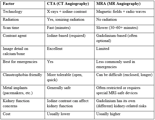
CTA vs MRA: Key Differences at a Glance
CT Angiography vs MRI Angiography: How Accuracy Compares
A common question patients ask is simply: which test finds problems better?
The honest answer is — it depends on what's being looked at. CTA is generally preferred over MRA for vascular imaging because it has faster acquisition time, higher spatial resolution, and a superior ability to detect calcifications, though MRA offers the advantage of avoiding radiation and iodinated contrast. For example, CTA has shown higher sensitivity and positive predictive value than MRA specifically for detecting intracranial artery narrowing.
That said, MRA isn't the "weaker" option across the board. MRA can actually be better than CTA at identifying a specific type of internal bleeding within the artery wall that occurs with cervical artery dissections. And in some vascular regions, the two tests perform almost identically. One meta-analysis looking at stenosis detection in dialysis access found no statistically significant difference in diagnostic performance between CTA and MRA, with both proving highly accurate.
Similarly, in imaging of blood vessels feeding tumors in the limbs, image quality between MRA and CTA showed no statistically significant difference, and MRA actually identified more feeding arteries and arteriovenous connections than CTA in that particular study.
The takeaway: neither scan is universally "more accurate." Accuracy depends heavily on the body part, the clinical question being asked, and even the specific scanning protocol used.
Radiation and Safety: The Biggest Differentiator
This is usually the deciding factor for many patients and doctors.
CTA uses ionizing radiation. A single scan carries a small, generally low risk, but it adds up if you need repeated imaging over months or years — which matters for people with chronic vascular conditions who get scanned regularly.
MRA avoids ionizing radiation entirely, making it the safer long-term option for patients who need frequent monitoring, children, and pregnant patients (when imaging is unavoidable).
On the contrast side, the safety profile flips depending on kidney function. MRA's gadolinium-based contrast carries a lower risk of kidney toxicity compared to CTA's iodine-based contrast, but gadolinium carries its own separate risk of a rare condition called nephrogenic systemic fibrosis in patients who already have significant kidney dysfunction. This is exactly why your doctor will usually check your kidney function (via a blood test) before ordering either scan if there's any concern.
Speed, Comfort, and Practical Considerations
Beyond accuracy and safety, real-world factors often tip the decision:
- Claustrophobia: MRI/MRA machines are long, narrow tubes, and scans can run 30 minutes or longer. CTA is a quick pass through a much shorter, more open scanner — often over in under 10 minutes.
- Metal in the body: Certain pacemakers, older aneurysm clips, and some other implants aren't compatible with MRI's strong magnetic field. In these cases, CTA becomes the default choice regardless of other factors.
- Motion sensitivity: CTA is less affected by motion artifacts, while MRA is more susceptible to motion artifacts as well as interference from magnetic field-incompatible devices. This makes CTA more forgiving for patients who have trouble holding still, including children or people in pain.
- Emergencies: In stroke centers and ERs, CTA is usually the go-to because it's faster and more widely available around the clock.
When Might Doctors Use Both?
Sometimes one scan alone doesn't provide the full picture. In complex cases, combining CTA and MRA can provide complementary information that may reduce the need for a more invasive procedure like digital subtraction angiography. This is especially true in complicated neurovascular or aneurysm cases where the two technologies catch different details.
So, CTA or MRA — Which One Should You Choose?
There's no single "better" scan — the right choice depends on your specific situation. Here's a simplified way to think about it:
CTA may be the better fit if:
- You need results quickly (emergency or urgent situation)
- You have a pacemaker or other MRI-incompatible implant
- You struggle with claustrophobia or can't stay still for long periods
- Your doctor needs to evaluate calcium deposits or bone-related vascular issues
- Cost or availability is a major factor
MRA may be the better fit if:
- You need to avoid radiation exposure (repeated monitoring, pregnancy, younger patients)
- You have reduced kidney function and iodine contrast is a concern
- Your doctor is investigating certain conditions, like arterial dissections, where MRA has an edge
- You have time for a longer scan and no MRI-incompatible implants
Ultimately, this decision is best made together with your physician or radiologist, who will weigh your medical history, the body part being examined, urgency, and your overall health profile. Both tests are safe, well-established, and capable of giving doctors the detailed vascular picture they need — the "right" one is simply the one that matches your circumstances.
Frequently Asked Questions
Is CTA or MRA more accurate?
Neither is universally more accurate. CTA tends to have an edge in speed, resolution, and detecting calcified plaque, while MRA can outperform CTA in specific scenarios like detecting arterial wall bleeding in dissections. Accuracy varies by the body region and condition being evaluated.
Does MRA use radiation?
No. MRA uses magnetic fields and radio waves, not X-rays, so it involves no ionizing radiation. CTA does use ionizing radiation.
Which is faster, CTA or MRA?
CTA is significantly faster, often completed in a matter of minutes. MRA typically takes 30 minutes or more.
Can I get an MRA if I have a pacemaker?
Not always. Many traditional pacemakers are not compatible with MRI machines. CTA is usually the safer alternative in these cases, though some newer "MRI-conditional" pacemakers may allow MRA under specific protocols.
Is CTA or MRA better for kidney patients?
It depends on the type and severity of kidney dysfunction, since both contrast agents (iodine for CTA, gadolinium for MRA) carry distinct risks. Your doctor will typically check kidney function before recommending either scan with contrast.
Do both scans require contrast dye?
CTA almost always requires iodine-based contrast. MRA can sometimes be performed without any contrast agent, depending on the vessels being examined and the scanning technique used.

Kate, Editorial Team at European Hospital & Healthcare Management, leverages her extensive background in Healthcare communication to craft insightful and accessible content. With a passion for translating complex Healthcare concepts, Kate contributes to the team's mission of delivering up-to-date and impactful information to the global Healthcare community.