
Photon-Counting & Dual-Energy CT: The Future of Angiography
Kate Williamson, Editorial Team, European Hospital & Healthcare Management
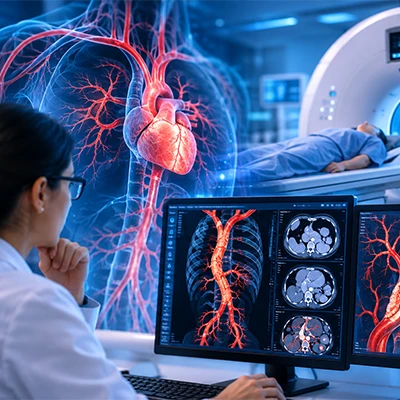
Photon-counting CT is transforming vascular imaging by capturing individual X-ray photon energies, delivering sharper resolution, lower radiation, and reduced contrast needs. Building on dual-energy CT's material differentiation strengths, this next-generation technology enables clearer plaque assessment and artifact reduction—positioning photon-counting CT angiography as the emerging standard for faster, safer diagnostic vascular imaging.
Introduction:
If you've spent any time around radiology circles lately, you've probably heard the buzz about photon counting CT angiography. It's not hype for hype's sake — this technology is genuinely reshaping how clinicians visualize blood vessels, catch disease earlier, and make decisions that used to require guesswork. Alongside its close counterpart, dual energy CT angiography, photon-counting detectors are quietly becoming the backbone of what many are now calling next-gen CT scanner vascular imaging.
Let's break down what's actually changing, why it matters, and what it means for patients and providers alike.
Why Traditional CT Angiography Started Showing Its Age
For decades, conventional CT scanners relied on energy-integrating detectors. These devices are reliable workhorses, but they have a fundamental limitation: they measure the total energy of X-ray photons hitting the detector, without distinguishing between individual photon energies. That means a lot of useful diagnostic information simply gets averaged out and lost.
This mattered most in angiography — the imaging of blood vessels — where subtle differences in tissue density, calcium buildup, and contrast enhancement can make or break a diagnosis. Radiologists have long dealt with issues like blooming artifacts from calcified plaques, beam hardening, and the need for higher contrast doses to get a clear picture. None of these problems are dealbreakers, but they add friction, cost, and occasionally diagnostic uncertainty to a process that's supposed to be precise.
Enter photon-counting detector (PCD) technology — a shift that's arguably the biggest leap in CT hardware since the introduction of multi-slice scanners.
What Makes Photon Counting CT Angiography Different
Photon-counting CT doesn't just measure the cumulative energy of incoming X-rays — it counts individual photons and sorts them by their specific energy levels in real time. Think of it like the difference between weighing a bag of mixed coins versus counting each coin and knowing exactly what denomination it is.
This photon-by-photon approach delivers a few game-changing benefits:
- Higher spatial resolution — because there's no need for scintillator crystals or light-guiding optics that blur signal, images come out sharper, which is critical when you're trying to assess a narrow, partially calcified coronary artery.
- Better contrast-to-noise ratio — vessels and soft tissue differentiate more clearly, even at lower radiation doses.
- Reduced electronic noise — because photon counting detectors don't need to convert light into an electrical signal (a step that introduces noise in traditional systems), the resulting scans are cleaner.
- Built-in spectral data with every scan — every single acquisition automatically carries multi-energy information, without needing a special dual-energy protocol.
That last point is subtle but huge. It means spectral imaging — once a niche, protocol-dependent feature — becomes the default, not the exception.
For clinicians managing complex vascular cases, this translates into being able to see through calcified plaque, better characterize stenosis severity, and reduce the iodine contrast load for patients with kidney concerns — all from a single acquisition.
Where Dual Energy CT Angiography Fits Into the Picture
Before photon-counting became clinically available, dual energy CT angiography was already pushing the boundaries of what a single scan could reveal. Dual-energy systems work by acquiring data at two different X-ray energy levels — typically using two separate tube-detector setups, rapid kilovoltage switching, or layered detectors.
The result is the ability to differentiate materials based on how they absorb X-rays at different energies. Iodine, calcium, and soft tissue all behave distinctly under different energy spectra, so dual-energy imaging can:
- Generate virtual non-contrast images from a single contrast-enhanced scan, cutting down on the need for a separate baseline scan (and the radiation dose that comes with it)
- Create iodine maps that highlight perfusion and enhancement patterns, useful for detecting pulmonary embolism, endoleaks after aneurysm repair, or subtle ischemia
- Improve plaque characterization, distinguishing calcified from non-calcified components in coronary and peripheral arteries
- Reduce metal and beam-hardening artifacts, which matters a lot for patients with stents, clips, or orthopedic hardware near the region of interest
In many ways, dual-energy CT laid the clinical and conceptual groundwork that photon-counting CT is now building on. The difference is that photon-counting doesn't need two separate energy acquisitions to get spectral data — it's inherently spectral, all the time, which simplifies workflow and often improves image quality further.
Next-Gen CT Scanner Vascular Imaging: What's Actually Changing at the Bedside
It's easy to get lost in the physics, so let's ground this in what actually happens in a clinic or hospital using next-gen CT scanner vascular imaging technology.
Lower radiation exposure. Photon-counting detectors are more dose-efficient because they don't waste signal on electronic noise. Some studies have reported meaningful dose reductions compared to conventional CT while maintaining or improving diagnostic image quality — a big deal for patients who need repeat vascular imaging over time, such as those being monitored after aortic aneurysm repair or stent placement.
Lower contrast volume requirements. Because spectral data allows for optimized "virtual monoenergetic" image reconstruction, radiologists can often achieve excellent vessel opacification with less iodinated contrast. That's meaningful for patients with chronic kidney disease or contrast allergies, who previously might have been poor candidates for CT angiography altogether.
Sharper small-vessel visualization. Coronary CT angiography has traditionally struggled with tiny, distal vessels and heavily calcified segments. Photon-counting's improved spatial resolution — sometimes down to submillimeter detail — helps radiologists assess these areas with far more confidence, potentially reducing unnecessary downstream testing like invasive catheter angiography.
Faster, simpler workflows. Since spectral data is captured by default, there's no need to plan ahead for a "dual-energy protocol." Technologists can scan first and decide later whether spectral analysis adds value, which streamlines scheduling and reduces protocol errors.
Better outcomes for complex patients. Patients with obesity, extensive vascular calcification, or metal implants — groups that have historically produced degraded CT images — tend to benefit disproportionately from these advances, since photon-counting and dual-energy techniques directly address the artifacts that plague these cases.
Real-World Applications Gaining Traction
Health systems investing in this technology are seeing applications expand quickly across specialties:
- Cardiology — coronary artery disease assessment, plaque characterization, and calcium scoring with improved accuracy
- Neurovascular imaging — detecting aneurysms, dissections, and stenosis with less artifact from bone and calcification
- Peripheral vascular disease — evaluating heavily calcified lower-extremity arteries in diabetic patients, a historically difficult population to image well
- Pulmonary embolism workups — iodine mapping helps confirm perfusion defects beyond just clot visualization
- Post-surgical vascular follow-up — monitoring stents, grafts, and aneurysm repairs with reduced metal artifact
Is This Technology Widely Available Yet?
Not universally — but adoption is accelerating. Photon-counting CT scanners received regulatory clearance in recent years and are gradually being installed in academic medical centers and major hospital systems, with broader community adoption expected to follow the typical technology diffusion curve seen with prior CT innovations. Dual-energy CT, by contrast, is already well established and widely available, making it the practical entry point into spectral imaging for many facilities not yet ready to invest in photon-counting hardware.
Frequently Asked Questions
What is photon counting CT angiography?
It's a vascular imaging technique that uses photon-counting detectors to individually register and energy-classify each X-ray photon, producing sharper images, better contrast resolution, and inherent spectral data with every scan — often at a lower radiation dose than conventional CT.
How is dual energy CT angiography different from photon-counting CT?
Dual-energy CT acquires images at two distinct energy levels using separate tube-detector configurations or rapid switching, while photon-counting CT captures the full energy spectrum in a single acquisition using specialized detectors. Both enable material differentiation, but photon-counting typically offers higher resolution and doesn't require special dual-energy protocol planning.
Does photon-counting CT reduce radiation dose?
Generally, yes. Because photon-counting detectors minimize electronic noise, they can often achieve diagnostic-quality images at lower radiation doses compared to traditional energy-integrating CT systems.
Is photon-counting CT better for patients with kidney disease?
It can be, since spectral reconstruction often allows for reduced iodinated contrast volumes while maintaining strong vessel opacification, which is beneficial for patients with contrast sensitivity or reduced kidney function.
Which hospitals offer next-gen CT scanner vascular imaging?
Availability varies by region, but adoption is growing fastest at academic medical centers, cardiovascular specialty hospitals, and large health systems investing in advanced imaging infrastructure. Checking directly with a local radiology department is the best way to confirm availability.
The Bottom Line
Photon-counting and dual-energy CT aren't just incremental upgrades — they represent a fundamental shift in how vascular imaging works. By capturing richer data with every scan, reducing radiation and contrast burden, and sharpening resolution in historically difficult cases, these technologies are pushing angiography toward a future that's faster, safer, and more precise. As more institutions adopt next-gen CT scanner vascular imaging systems, patients and clinicians alike stand to benefit from diagnostic clarity that simply wasn't possible a decade ago.
The shift is already underway — the question now isn't if photon-counting becomes the new standard, but how quickly.

Kate, Editorial Team at European Hospital & Healthcare Management, leverages her extensive background in Healthcare communication to craft insightful and accessible content. With a passion for translating complex Healthcare concepts, Kate contributes to the team's mission of delivering up-to-date and impactful information to the global Healthcare community.