Advancing Thrombosis Care through Patient-Centered Approaches
Eirik Tjønnfjord, Senior Consultant, Kalnes Hopsital and Rikshospitalet
AI and Machine learning is great new tools that have taken the world with storm over the last years. It has also found its place in medicine. I believe that these innovative tools will make the medicine better used in the right hands and with a critical view. At my work at the department of thrombosis and coagulation we use patient centered tools to improve the diagnosis and streamline the patient care.
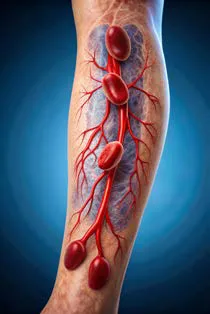
In my work at the Department of Thrombosis and Hemostasis, we frequently see patients referred to the hospital with suspected DVT (deep venous thrombosis) or PE (pulmonary embolism). Unfortunately, they often end up waiting several hours – or even have to return the next day – for a leg ultrasound or a CT scan of the lungs.
This is particularly frustrating because DVT patients could be diagnosed and treated much more quickly if the physicians or nurses in the ER could perform ultrasounds themselves, without having to wait for the radiologists. This becomes even more critical after normal working hours or on weekends, where faster diagnoses could save valuable time and reduce hospitalization.
At a partner hospital, they’ve implemented an education program where ER doctors are trained to perform ultrasounds. This initiative has reduced the time to diagnosis from 8 hours to just 2, allowing them to start treatment and discharge patients much faster. The benefits are clear: it saves time for doctors and patients, ensures quicker and more accurate treatment, and cuts down on hospitalization days and costs. Plus, it frees up the radiology department to focus on other important exams like CTs and MRIs.
If we could share this knowledge and expertise across all hospitals, we could help more patients, save time, reduce costs, and optimize space.
Artificial Intelligence (AI) and machine learning (ML) are advancing rapidly, and they offer incredible potential to optimize treatment and save time in a busy hospital setting. Combined with a patient-centered approach to diagnosis, these technologies are becoming essential as our population grows and people expect faster answers to their health concerns.
In today’s fast-paced healthcare environment, where everyone is working hard to provide the best possible care, it’s crucial to explore ways to improve and speed up diagnostic tools and decision-making processes. The way things have always been done needs to be put under the microscope, critically reviewed, and updated to meet the demands of the future. What was cutting-edge in 2023 might be outdated by 2024 and practically ancient by 2025. This also raises questions about who performs various tasks in hospitals – can we delegate certain responsibilities to other specialties or healthcare workers, such as nurses or paramedics?
In my role as a hematologist with a special focus on thrombosis and hemostasis, I’ve worked closely with other medical fields to ensure accurate diagnoses. This often involves collaboration with radiologists for ultrasounds and CT scans, cardiologists for pulmonary embolism cases, and general practitioners (GPs) for follow-up care.
With an aging population and increasing pressure to deliver quick and accurate diagnoses, the radiology department, like many others, is feeling the strain. CT, MR, and PET scans are in higher demand than ever, especially for cancer diagnosis and monitoring, and these procedures take up a significant amount of time due to their life-changing implications.
However, deep vein thrombosis (DVT) is another condition that must be taken seriously, as it can lead to more severe complications like pulmonary embolism if not recognized and treated promptly. In our busy emergency department, we’ve seen patients referred to the ER with suspected DVT who can wait up to 8-12 hours before an ultrasound is performed. While they are usually started on anticoagulation and sent home, with instructions to return the next day, this isn’t always practical – especially for older patients or those who live far away. If they have to stay overnight, they occupy a bed that could be used for more critically ill patients.
To address these challenges, we've launched a program to train emergency doctors to perform these ultrasound scans themselves. Their results are compared to the gold standard set by radiologists and ultrasound technicians, and the outcomes have been impressive, even after a relatively short training period. The process is thorough: the ER doctors perform ultrasounds, which are then cross-checked by radiologists multiple times before the doctors are allowed to work independently.
Typically, an ER doctor will do 15-20 ultrasounds, after which a radiologist will perform the same scans without knowing the ER doctor’s results. If the ER doctor’s results match the radiologist's findings across all cases, the doctor then conducts another 15-20 ultrasounds independently, with the results still being verified by radiologists. Once they consistently achieve accurate results, they’re certified to perform DVT ultrasound investigations on their own.
With this new approach, patients can be examined, diagnosed, treated, and safely sent home within 2-4 hours. This is a significant improvement compared to the traditional process, where patients could wait anywhere from 6 hours to overnight. Sometimes, they were even sent home with instructions to return the next day, having waited for blood tests, vitals, and examinations by multiple healthcare providers before being placed on potentially risky medications.
New ER doctors are continuously trained by experienced colleagues, ensuring that they stay up-to-date while new trainees are brought up to speed. This approach not only saves time and effort but also streamlines the diagnosis and treatment process, ultimately saving money for the hospital and enabling us to help more patients.
Studies have shown that with practice, ER doctors can perform ultrasounds just as accurately as radiologists or ultrasound technicians. In other hospitals in Norway, like Skien, they’re even training acute care nurses to perform ultrasounds in the ER. This frees up ER doctors to focus on more critically ill patients, while nurses can quickly check ultrasound results and prescribe anticoagulation if needed.
These initiatives are win-win situations: hospitals save money, reduce hospitalizations, and speed up patient care, while radiologists can focus on more urgent cases. Perhaps most importantly, young doctors and healthcare workers, like nurses, develop new skills, boosting their confidence and motivation to continue working in what can be a demanding and challenging environment. This continuous learning and skill development help prevent burnout and keep talented professionals engaged in their work.
So far, this program has been a great success at our hospital. It took some time to set up the education process and get the first group of doctors trained, but now things are moving much faster. Before long, experienced ER doctors can even join the training and education team, helping to bring new doctors up to speed.
One key lesson we’ve learned is the importance of ensuring that the doctors, nurses, and other healthcare workers involved have a genuine interest in learning and working with ultrasound. It’s essential for them to stay engaged with ultrasound regularly to keep their skills sharp. While I firmly believe – through my work with my partner at NorVue, where we create educational ultrasound videos – that anyone can learn to perform bedside ultrasound, it requires dedication. In my opinion, checking for DVT is one of the easiest ultrasound skills to learn, but becoming confident takes endurance, patience, and persistence. And to truly excel, it requires repetition and hands-on experience. Some of the doctors who were trained but don’t use ultrasound daily tend to lose that "feel" for it – they don’t have it in their fingertips anymore.
This is just one example of how point-of-care diagnostics can be incredibly useful in a busy hospital environment, but there are many others. For instance, surgical nurses could be trained to interpret X-rays for fractures, as seen in other institutions. At Akershus University Hospital, nurses perform bone marrow aspirations, a task that’s also done by medical students at Odense University Hospital in Denmark. General practitioners (GPs) can even be trained to perform ultrasounds, so patients don’t always have to come to the hospital at all.
The pace of healthcare is accelerating, and patients increasingly expect quick diagnoses and treatments. Many have already turned to Google or other sources and might come in with their own ideas about what’s wrong. To keep up with these expectations and to help more people in less time, we need to develop new skills, delegate tasks where possible, and collaborate across specialties. By sharing knowledge and working together, we can better prepare for a future with a growing, longer-living population that demands more from our healthcare systems. Ultimately, we’re only as strong as the weakest link in our team, but by pulling together, we’re much more likely to succeed.
Reference list:
1. Hercs D. et al. Ultrasound Perfromed by Emergency Physicians for Deep Vein Thrombosis: A systematic review. Western Journal of Emergency Medicine. Volume 25, No 2;March 2024. 282-290.
2. She hunts for bloodcloths. Sykepleien. 14.06.2024 (Norwegian)
3. Anthony Summers at al. Can nurses interpret X-rays safely without formal tuition?. Accident and Emergency Nursing: Volume 13, Issue 3, July 2005, 162-166
4. Lasse Johan is a nurse and do bonemarrow biopsy. Sykepleien. 11.06.2023 (Norwegian)
Author Bio
Eirik Tjønnfjord, a senior consultant at Kalnes Hospital and Rikshospitalet in Norway, specializes in hematology with a particular passion for benign hematology and CLL, and leads several related studies. His work focuses on thrombosis and coagulation, and he has a special interest in rare conditions such as PNH and CAD. In addition to his clinical practice, he uses ultrasound in his work and, together with his wife, runs Norvue, an ultrasound company that teaches health workers.